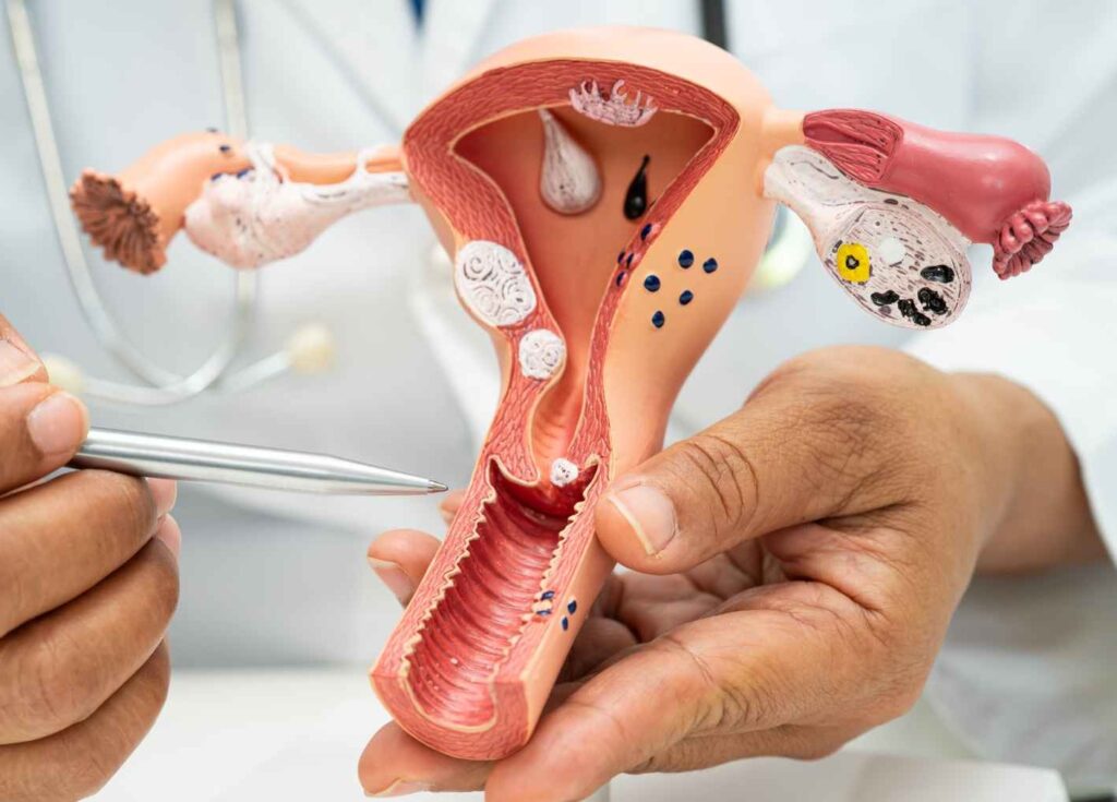
Contemporary women have fewer children, have them later in life, and spend a much larger proportion of their adult years without pregnancy. The demographic transformation is evident. The question that is beginning to interest geroscience is far less obvious: Could this shift in reproductive trajectory have consequences for longevity and, above all, for the years lived in good health?
The question takes on a new dimension with the so-called Reproductive Resilience Hypothesis, proposed by researchers at the Buck Institute for Research on Aging and published as a perspective article in Cell. Their approach does not argue that having children lengthens life. It proposes something more sophisticated: evolution may have developed maintenance and resilience mechanisms in females precisely because their survival matters for offspring and for future reproductive success.
The article suggests that pregnancy, lactation, reproduction, menopause, and ovarian function should be studied as relevant variables in the biology of aging, not merely as reproductive events.
The hypothesis is not yet proven. What is interesting is that, when tested against available clinical and epidemiological research, some correspondences emerge, along with results that call for nuance. The evidence does not paint a linear relationship between reproduction and longevity, and that is precisely where one of the most intriguing questions for current research lies.
Having children is not a decisive variable
Studies attempting to link number of children and longevity do not offer a simple answer.
A meta-analysis of 18 studies with more than 8 million participants found a non-linear association between parity and mortality. People without children showed higher overall mortality than those who had at least one child, while the lowest risk appeared with moderate parity. The point of lowest mortality was around three or four births. Beyond high parity levels, the advantage disappeared. (pmc.ncbi.nlm.nih.gov)
Another prospective meta-analysis, focused on cardiovascular mortality and including nearly one million participants, also found a non-linear relationship: the greatest risk reduction appeared around four births, although the comparison between women with children and women without children did not reach conventional statistical significance. The authors themselves noted considerable heterogeneity across studies. (pubmed.ncbi.nlm.nih.gov)
The data, therefore, do not allow us to claim that the more children a woman has, the longer she will live. Nor do they show that having no children is harmful to health. What appears in some large studies is a statistical association with low or moderate parity, although numerous social, economic, behavioral, and prior health factors may lie behind it, making it difficult to establish a causal relationship.
The available evidence forces us to separate reproduction as a biological experience from motherhood as a social phenomenon. Having one, three, or five children involves different physiological trajectories, but also personal, economic, and cultural circumstances that can independently influence health and survival.
If developed societies are moving from three or four children per woman to one or two, we might ask whether we are hypothetically eliminating a possible biological advantage. We have no evidence to say so.
Available data do not allow us to calculate how much declining fertility might affect a population's longevity. The fundamental reason is that the number of children does not by itself determine the duration of ovarian function.
A woman can have a single child and reach menopause at 53. Another can have four children and experience menopause at 40. From the perspective of biological aging, those two reproductive histories are completely different.
Longevity research therefore needs to look beyond parity. If we want to understand the relationship between reproduction and aging, it is probably more useful to study how long the reproductive system remains functional than to simply count how many pregnancies a woman has had.
The length of reproductive life seems to matter more
Reproductive life roughly spans the interval between menarche and menopause. A large international collaboration that pooled individual data from more than 300,000 women found a clear relationship between a short reproductive life and higher cardiovascular risk. Women with fewer than 30 years of reproductive life had a substantially higher risk of cardiovascular disease than those with a duration of 36–38 years.
A significant portion of that relationship disappeared when accounting for age at menopause, a finding that places the menopausal transition at the center of the observed association.
The signal reappears in more recent research on mortality. In an analysis of women over 65, a longer reproductive life was associated with lower overall and cardiovascular mortality.
A study published in 2026 adds an even more intriguing piece. In more than a thousand American women over 50, later menopause and a longer reproductive life were associated with a slowdown in certain biological aging indicators measured via epigenetic clocks. (nature.com)
Here, a possible correspondence with the Reproductive Resilience Hypothesis emerges. The relevant variable would not necessarily be pregnancy itself, but rather the capacity of the female organism to maintain healthy reproductive function for a longer period.
The difference is important. Maintaining ovarian function for more years may be a consequence of greater overall biological resilience and, at the same time, contribute to maintaining certain physiological systems. Research has not yet established which part corresponds to each causal direction.
Early menopause poses the inverse problem
The evidence on early menopause is considerably more consistent.
Women who experience premature or early menopause show, in numerous studies, a higher subsequent risk of cardiovascular disease, metabolic alterations, and mortality.
The interpretation should not be reduced to a single explanation either. Early menopause may be related to genetic, autoimmune, metabolic, toxic, surgical, or environmental factors. It can also function as a marker of an organism exhibiting certain characteristics of accelerated aging.
Epigenetic research is beginning to reinforce this second possibility. DNA methylation clocks allow for estimating a biological age that does not always coincide with chronological age, and some studies have found associations between early menopause and epigenetic acceleration, although the magnitudes and mechanisms are still being investigated.
The 2026 research cited above is especially relevant because it finds the relationship in the opposite direction: later menopause and a longer reproductive lifespan were associated with less acceleration of certain epigenetic clocks. (nature.com)
The question therefore ceases to be whether menopause “causes” aging. Scientific interest shifts toward the possibility that aging of the reproductive system constitutes one of the most visible indicators of the rate at which the female organism is aging.
Recent research introduces an element that prevents building too comfortable a narrative around reproduction.
The 2026 study found that a higher frequency of pregnancies was associated with small increases in some epigenetic age indicators. Women with five or more pregnancies showed greater odds of acceleration in certain clocks, and five or more births also appeared associated with certain biological aging markers. (nature.com)
The result fits with previous research.
A study published in 2019 found weak associations between number of births and epigenetic age. The associations weakened after adjusting for body mass index. (pubmed.ncbi.nlm.nih.gov)
A study of Scientific Reports conducted with 4,418 women also found a U-shaped relationship between parity and biological age acceleration in postmenopausal women. The lowest levels of acceleration appeared among those who had had three or four children. (nature.com)
The overall set of results allows us to propose that reproduction may simultaneously present physiological costs and resilience mechanisms. Pregnancy involves an enormous cardiovascular, immune, metabolic, and endocrine remodeling, while lactation adds another energy demand. Repeating these processes may carry a cumulative cost. The ability to go through them and subsequently maintain prolonged ovarian function could, in turn, be a signal of resilience.
There is not necessarily a contradiction between both observations. Both things may be happening at the same time.
A woman may assume a reproductive cost and, at the same time, show greater resilience
This apparent paradox is central to interpreting the evolutionary hypothesis.
The classic model of the disposable soma proposes that resources allocated to reproduction cannot simultaneously be devoted to bodily maintenance. The Reproductive Resilience Hypothesis proposes that this explanation may be incomplete. In certain species, natural selection may have favored mechanisms capable of offsetting part of that cost when the mother's survival remains important for the offspring.
The article by Cell insists that the number of reproductions is not the fundamental issue. What would be decisive is whether continued survival still holds value for reproductive success, offspring care, or inclusive fitness.
Translated to humans, this opens a particularly interesting hypothesis: reproduction could impose a physiological cost and, at the same time, select for or reveal individuals with greater maintenance and repair capacity.
Having children would not necessarily be the mechanism that produces longevity. It could be, in certain cases, an event that allows observing an organism's resilience.
The Finnish twin study adds another piece
A study published in Nature Communications in 2025 examined the relationship between reproductive history, epigenetic aging, and survival in the Finnish Twin Cohort.
The researchers observed a higher mortality risk among childless women and also among those with five or more children, using women with three children as the reference. Specifically, mortality risk was 37% higher in childless women and 22% higher in those with five or more children compared to the three-child group, according to the model presented by the authors. (nature.com)
The shape of the association is therefore again approximately a U. The research does not appear to support a biological rule whereby having many children protects against aging, just as it does not allow establishing a universal penalty associated with having none.
The intermediate zone is what appears associated with better outcomes in several studies, although even these data must be interpreted with caution. Women who have no children constitute an extraordinarily heterogeneous group. Infertility, the voluntary decision not to have offspring, economic situation, marital status, prior health, and social circumstances can lead to the same reproductive outcome through completely different pathways.
What does this mean for societies that are having fewer and fewer children?
It is worth separating demography from individual biology.
The fact that a society has a low fertility rate does not automatically mean that its women will age worse. A population can have fewer births and, simultaneously, women with a long reproductive lifespan, relatively late menopause, better cardiovascular control, less smoking, more physical activity, and better healthcare access. From the standpoint of healthy longevity, those factors may be far more important than going from two children to one.
Current scientific evidence also provides no basis for recommending having more children as a longevity strategy. Such a recommendation would mean turning epidemiological associations into causality, something the available studies do not allow.
Demographic transformation does raise an interesting scientific question: societies with later motherhood are modifying the temporal relationship between reproduction, aging, and menopause.
A woman who has her first child at 38 and a woman who has one at 24 do not follow exactly the same biological trajectory. The former may be closer to the perimenopausal transition during her final reproductive years, while the latter has more time between pregnancy and reproductive aging. This difference does not mean that one will necessarily live longer than the other; it shows that reproductive timing can carry its own significance.
Modern demographics have reduced the number of pregnancies and shifted motherhood to later ages, while the age of menopause has not increased at the same rate.
This situation creates a distinct biological trajectory. Women may spend more years of their adult lives without reproducing, while continuing to experience an ovarian trajectory determined by the progressive loss of follicles and the subsequent menopausal transition.
That is why, for longevity research, number of children, number of pregnancies, age at first birth, age at last birth, age at menopause, and length of reproductive lifespan should be analyzed separately.
Grouping all these factors under the word “reproduction” can obscure precisely the mechanism we are looking for.
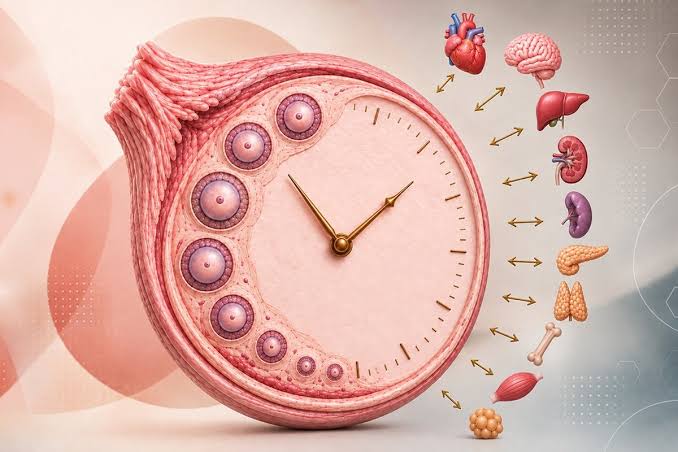
The ovary as a biological clock
This is probably the most provocative part of the entire discussion.
The ovary produces estradiol, progesterone, and other mediators, but its activity is not limited to classic sex hormones. There is ongoing communication with metabolism, bone, the cardiovascular system, the brain, and the immune system.
The original article proposes precisely interpreting the ovary as a kind of physiological coordination center. Its authors warn, however, that its decline interacts with many other aging mechanisms: chronic inflammation, mitochondrial dysfunction, genomic instability, stem cell exhaustion, and tissue damage.
The hypothesis that emerges is far more interesting than claiming that “ovaries make you live longer.” There could be a bidirectional relationship: an organism that ages more slowly preserves its ovarian function for longer, and preserved ovarian function may help maintain certain bodily systems.
Determining how much weight each direction carries is one of the great pending challenges.
Research on female longevity has tended to study menopause as an event that occurs mid-life and subsequently increases certain risks. The new perspective proposes looking backward and studying the years before menopause, the age at which ovarian reserve declines, the AMH trajectory, age at first and last pregnancy, metabolic complications of pregnancy, the duration of reproductive life, and finally, the speed at which each woman goes through the menopausal transition.
This approach would allow a shift from medicine based exclusively on chronological age to medicine that also considers reproductive age as a dimension of female biological aging.
Available evidence does not show that lower birth rates will reduce healthy longevity in women, nor the opposite. Nor does it show that having multiple children protects against aging.
What consistently emerges is a much more complex pattern:
- Low or moderate parity appears in various studies to be associated with lower mortality than nulliparity or very high parity. (pmc.ncbi.nlm.nih.gov)
- High parity, especially five or more births, appears in some recent studies to be associated with certain indicators of biological aging. (nature.com)
- A longer reproductive life is linked to better cardiovascular outcomes and, in recent research, to slower acceleration of some epigenetic clocks. (nature.com)
- Premature or early menopause is associated with higher cardiovascular risk and mortality in numerous studies.
- The number of children, by itself, seems to be a much less precise indicator than the duration and quality of reproductive function.
The most prudent conclusion is also the most interesting: female longevity appears to be related to the reproductive trajectory, but not necessarily to the number of children.
A new frontier for female longevity medicine
If this line of research is confirmed, preventive medicine may need to pay much more attention to each woman's reproductive history. Not to recommend pregnancies, nor to artificially delay menopause without knowing its risks and benefits, and not to turn the age of menopause into a verdict on a woman's future.
The usefulness would lie elsewhere: identifying women who experience especially early reproductive loss and studying whether that signal can be used to anticipate cardiovascular, metabolic, bone, neurological, or systemic aging risk.
The Reproductive Resilience Hypothesis itself proposes precisely that reproductive history should be incorporated into aging studies and that biomarkers should consider reproductive status, not just chronological age.
This perspective could change the way we understand menopause. Not as an isolated date or simply the end of fertility, but as a signal within a biological trajectory that began decades earlier.
For geroscience, what matters most is: how long an organism managed to maintain a metabolically competent reproductive system and what was happening, at the same time, in its heart, brain, bones, mitochondria, and immune system.
Science does not yet have the definitive answer. The direction of research is becoming increasingly clear, however: to understand how women age, we will need to study much better how their ovaries age. That also changes the question about longevity. Perhaps the future of female preventive medicine does not lie in merely measuring how old a woman is, how many children she has had, or how long she has been in menopause. We may need to know how fast she has traveled her own reproductive trajectory and what relationship that trajectory has with the aging of the rest of her body.